
Acceso de los editores
Heart Valve Disease
Eingesendet von. Dr. Carlos Porras Der Artikel wurde in Cirugía Cardiaca, Cardiología Clínica publiziert und mit den Tags treatment, valve, mitral, aortic, stenosis, regurgitation, surgery versehen


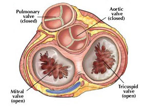
Every normal heart consists of heart muscle and four valves. Two valves are located between forechambers (atria) and pumping chambers (ventricles) as inlet valves, the other two are between ventricles and big arteries (aorta, pulmonary artery) as outlet valves of the pumping chambers. These valves regulate inflow into the heart and outflow into lungs of the body through coordinated opening and closing.
A disease may be caused by an inborn anomaly that gets worse with age. Some heart valve disease may only occur with wear due to increasing age or as a consequence of infections with bacteria.
Signs of heart valve disease may vary. The disease is often unrecognized for many years because the heart compensates the functional disturbance and patient does not experience any symptoms. If symptoms occur, decreased exercise tolerance is the most frequent. This may be felt as shortness of breath on physical exertion, increased fatigue, or simply “getting slower”. Chest pain may occur with some diseases, sometimes swelling of the ankles is noted.

The valves that are most frequently affected are those of the left heart. The mitral valve is the inlet valve of the left ventricle, and the aortic valve the outlet valve. One can differentiate between two principal functional changes, narrowing during outflow (stenosis) or leak during the closed phase (regurgitation). Sometimes a combination of the 2 disturbances may exist. The exact type of valve dysfunction may be detected by ultrasound (echocardiogram). Once a certain level of functional impairment is reached medication is not sufficient to stabilize the heart, and an operation or intervention is necessary. Without surgery principally the situation will continue to get worse, and ultimately additional damage to the heart muscle will develop that will contribute to heart failure or death.
|
|


- Avenida de los Argonautas, s/n, 29630, Benalmádena, (Málaga)
- Telefonnummer:
(+34) 952 367 190
Gemeinsam sind wir stark
Das Institut für Kardiotechnik vereint das Know-how aus vier Klinikbereichen, um erstklassige kardiologische Behandlungsleistungen zu erbringen.
ICTA: als Team am Puls der Zeit.
Los más leídos
- ¿Qué esperar tras su operación de corazón? (96208 hits)
- Estoy en tratamiento para la hipertensión arterial. ¿Son fiables los aparatos que miden la presión arterial de forma automática? (66252 hits)
- Aneurismas de aorta ascendente: ¿cuándo hay que operar? (56390 hits)
- Insuficiencia aórtica severa: ¿cuándo hay que operar? (39163 hits)
- Evaluacion de los antiinflamatorios en el riesgo cardiovascular (Diclofenaco, Ibuprofeno y Naproxeno) (37150 hits)
- Me voy a operar del corazón. Preguntas frecuentes (FAQ) (35848 hits)
- ¿Qué debe saber si se va a operar del corazón? (34759 hits)
- ¿El consumo moderado de alcohol es bueno para el corazón? (34372 hits)
- Valvulopatías (enfermedad valvular cardiaca): Generalidades (33453 hits)
- El sexo y las enfermedades cardiacas: ¿es seguro el sexo en pacientes con enfermedades del corazón? (31759 hits)
- Trucos para pacientes en autocontrol de tratamiento anticoagulante con Sintron o Warfarina (27625 hits)
- Tratamiento médico de la insuficiencia aórtica (26903 hits)
Málaga Health

Marfan-Kanal
Das Ärzteteam des Andalusischen Instituts für Kardiotechnik pflegt die in spanischer Sprache vorbildhafte Website zum Marfan-Syndrom.


Nube de etiquetas
Arrhythmien
Auf der Arrhythmien-Station der Herz-Abteilungp behandeln wir Patienten mit Herzrhythmusstörungen. Die Station setzt sich aus zwei spezifischen Bereichen zusammen.
Marfan-Station
Multidisziplinäres Team zur Diagnose, Kontrolle und Behandlung von Patienten mit Marfan-Syndrom.
Herzchirurgie
Die Behandlungstechniken moderner Herzchirurgie: Koronarchirurgie mit und ohne extrakorporalen Kreislauf, konventionelle Herzklappenchirurgie, usw.
Aortenklappen-Station
Pionierteam in der Aortenklappenrekonstruktion. Wir führen Eingriffe an Aortenwurzelaneurysmen in perfekt ausgestatteten Operationssäulen durch.
Klinische Kardiologie
Elektrokardiogramm (EKG), Ergometrie und Echokardiogramm: Praxen in Benalmádena (Krankenhaus Hospital Xanit) und in Fuengirola.
Plötzlicher-Herztod-Station
Früherkennung der Ursachen von Koronarerkrankungen, die zum plötzlichen Herztod beim Sport führen können.
Hämodynamik
Koronarangiographie, Untersuchung von Patienten mit Herzklappenerkrankung:: angeborene Herzfehler beim Erwachsenen, transseptale Katheterisierung, usw.
Kardiologische Rehabilitation
Fachservice zur vollständigen Rehabilitation von Patienten nach An gi na Pec to ris, Herzinfarkt, Operationen, usw.
| Kontakt: 952 367 190 | Rechtshinweis | Sitemap |

![]() Herzchirurgie Málaga, Kardiologie en Málaga, Kardiologen Málaga, Arrhythmien, Katheterisierung, Herzchirurg Málaga, Herzklappenchirurgie, Behandlung des Marfan-Syndroms, Herzchirurgie in Málaga, motorische Rehabilitation, kardiologische Rehabilitation, Aortenklappenrekonstruktion, Kardiologe Costa del Sol.
Herzchirurgie Málaga, Kardiologie en Málaga, Kardiologen Málaga, Arrhythmien, Katheterisierung, Herzchirurg Málaga, Herzklappenchirurgie, Behandlung des Marfan-Syndroms, Herzchirurgie in Málaga, motorische Rehabilitation, kardiologische Rehabilitation, Aortenklappenrekonstruktion, Kardiologe Costa del Sol.