Acceso de los editores
Heart Valve Disease
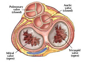
Опубликовал. Dr. Carlos Porras Данная статья была опубликована в Cirugía Cardiaca, Cardiología Clínica и с тегами treatment, valve, mitral, aortic, stenosis, regurgitation, surgeryEvery normal heart consists of heart muscle and four valves. Two valves are located between forechambers (atria) and pumping chambers (ventricles) as inlet valves, the other two are between ventricles and big arteries (aorta, pulmonary artery) as outlet valves of the pumping chambers. These valves regulate inflow into the heart and outflow into lungs of the body through coordinated opening and closing.
A disease may be caused by an inborn anomaly that gets worse with age. Some heart valve disease may only occur with wear due to increasing age or as a consequence of infections with bacteria.
Signs of heart valve disease may vary. The disease is often unrecognized for many years because the heart compensates the functional disturbance and patient does not experience any symptoms. If symptoms occur, decreased exercise tolerance is the most frequent. This may be felt as shortness of breath on physical exertion, increased fatigue, or simply “getting slower”. Chest pain may occur with some diseases, sometimes swelling of the ankles is noted.
The valves that are most frequently affected are those of the left heart. The mitral valve is the inlet valve of the left ventricle, and the aortic valve the outlet valve. One can differentiate between two principal functional changes, narrowing during outflow (stenosis) or leak during the closed phase (regurgitation). Sometimes a combination of the 2 disturbances may exist. The exact type of valve dysfunction may be detected by ultrasound (echocardiogram). Once a certain level of functional impairment is reached medication is not sufficient to stabilize the heart, and an operation or intervention is necessary. Without surgery principally the situation will continue to get worse, and ultimately additional damage to the heart muscle will develop that will contribute to heart failure or death.
|
|
Эффективность совместной работы

В Институте высокотехнологичной кардиологии знания и опыт специалистов четырех направлений сводятся воедино для предоставления услуг высочайшего класса по диагностике и лечению кардиологических заболеваний.
ICTA: совместные усилия сплоченного коллектива специалистов
Los más leídos
- ¿Qué esperar tras su operación de corazón? (91134 hits)
- Estoy en tratamiento para la hipertensión arterial. ¿Son fiables los aparatos que miden la presión arterial de forma automática? (62819 hits)
- Aneurismas de aorta ascendente: ¿cuándo hay que operar? (53434 hits)
- Insuficiencia aórtica severa: ¿cuándo hay que operar? (36492 hits)
- Evaluacion de los antiinflamatorios en el riesgo cardiovascular (Diclofenaco, Ibuprofeno y Naproxeno) (33312 hits)
- Me voy a operar del corazón. Preguntas frecuentes (FAQ) (32968 hits)
- ¿Qué debe saber si se va a operar del corazón? (31908 hits)
- ¿El consumo moderado de alcohol es bueno para el corazón? (30879 hits)
- Valvulopatías (enfermedad valvular cardiaca): Generalidades (30214 hits)
- El sexo y las enfermedades cardiacas: ¿es seguro el sexo en pacientes con enfermedades del corazón? (27809 hits)
- Trucos para pacientes en autocontrol de tratamiento anticoagulante con Sintron o Warfarina (24603 hits)
- Tratamiento médico de la insuficiencia aórtica (24239 hits)
Nube de etiquetas
Аритмии
В отделении аритмий кардиоцентра мы помогаем пациентам с различными нарушениями ритма сердца. В нем имеется два подразделения.
Отделение синдрома Марфана
Многопрофильный коллектив специалистов, выполняющий диагностику, диспансерное наблюдение и лечение пациентов с синдромом Марфана.
Кардиохирургия
Лечение методами современной кардиохирургии: аортокоронарное шунтирование с применением и без применения искусственного кровообращения, традиционное протезирование клапанов сердца и т. д.
Отделение проблем аортального клапана
Наши специалисты ведут новаторскую работу по реконструкции аортального клапана. Мы выполняем хирургическое лечение аневризмы корня аорты в прекрасно оснащенной операционной.
Клиническая кардиология
Электрокардиограммы (ЭКГ), эргометрия и эхокардиография (УЗИ сердца): обследование и консультации в Бенальмадене (больница Xanit) и во Фуэнхироле.
Отделение внезапной сердечной смерти
Раннее выявление причин атеросклероза коронарных артерий, который может привести к внезапной смерти спортсменов.
Нарушения гемодинамики
Коронарография, обследование пациентов с нарушением работы сердечных клапанов: : врожденные пороки сердца у взрослых, транссептальная катетеризация и т. д.
Кардиореабилитация
Специализированная служба комплексной реабилитации пациентов, перенесших стенокардию, инфаркт миокарда, находящихся в послеоперационном периоде
и т. д.


